Refer a Patient
 Pay Bill
 Patient Portal
 (800) 714-2020
What Do Glaucoma Test Results Mean?
Glaucoma is one of the leading causes of blindness in the U.S., which is partially because this condition goes asymptomatic until its late stages. However, this staggering statistic also stems from a lack of eye health maintenance among those most likely to develop glaucoma. Regular and thorough exams are needed to ensure that you are not developing this disease and to catch it early enough to slow its progression and reduce vision loss – but the process can be confusing.
Continue reading for the basics of various glaucoma tests and how to understand the test results with the help of the top eye doctors in greater Dallas.
Types of Glaucoma Tests
If you are over 40, have a family history of glaucoma, or have conditions that make you more susceptible to it, like diabetes or sleep apnea, then you should get tested at least yearly. Your eye doctor may use any combination of the six standard glaucoma tests to give you a proper diagnosis.
Why are there so many glaucoma tests? This condition can be hard to catch, especially since having any particular characteristic may not mean you absolutely have it.
Tonometry (Inner Eye Pressure)
Tonometry gauges your eye pressure as a potential sign of glaucoma. After numbing your eyes, your doctor will use a tonometer to measure your intraocular pressure (IOP).
The normal range for IOP is typically 12-22mm Hg, while indications of glaucoma generally begin at the 20mm point. However, it is still possible that someone with a “normal” reading can have glaucoma.
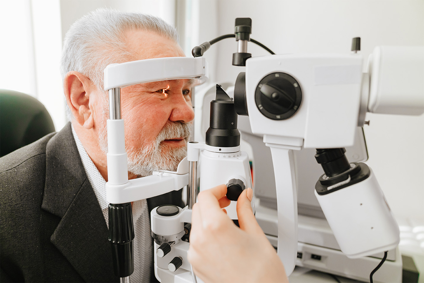
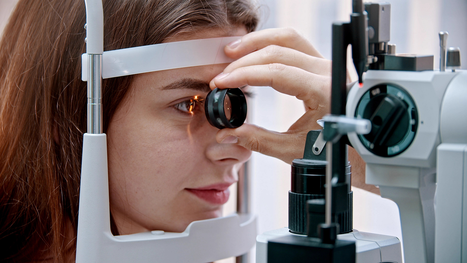
Ophthalmoscopy (Appearance of the Optic Nerve)
Ophthalmoscopy looks for optic nerve damage or abnormalities. Your pupils are first dilated to make the shape and color of the optic nerve more visible. Using a biomicroscope, your eye doctor magnifies and illuminates the area to view its appearance.
A cupped shape and non-pink color can signal abnormalities requiring further testing.
If the optic nerve’s eye pressure and appearance are cause for concern, then the best eye doctors in greater Dallas will conduct a series of the following tests to give a formal diagnosis.
Perimetry (Field of Vision)
To determine whether your vision has been affected and to what extent, your eye doctor will conduct a perimetry test or a visual field test to get a map of your complete visual range. Using one eye at a time, you will look at a central focal point and be asked to identify when different light spots come into view in your peripherals.
Don’t worry if you experience delays in your blind spots, as this is totally normal. Remain relaxed and answer accurately to ensure that you receive the proper treatment. After diagnosis, your doctor will run a perimetry test 1 to 2 times a year to keep an eye on your vision.
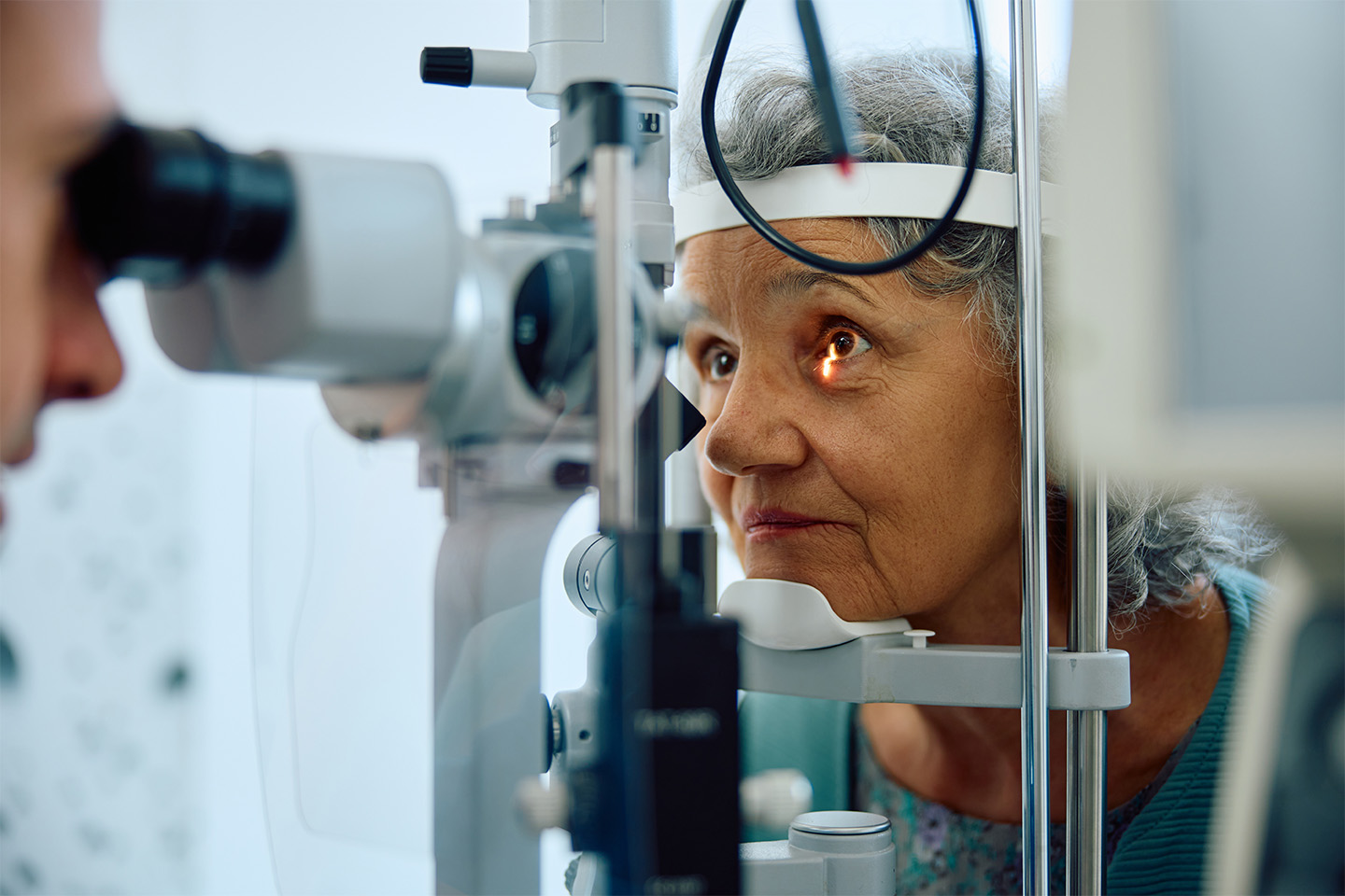
Gonioscopy (Angle at the Iris & Cornea)
The gonioscopy takes a deeper look at the drainage system and helps determine which form of glaucoma is present in the eye. A dysfunctional system can cause increased eye pressure and degrade the optic nerve, leading to glaucoma. To view this, your eye doctor will administer numbing drops, then use a special handheld mirrored lens to view the angle where the iris meets the cornea.
If this angle is wide and open, it is considered the more common Primary Open-Angle glaucoma. If it is narrow and closed, it is acute or narrow-angle, which is more medically severe.
Pachymetry (Corneal Thickness)
Corneal thickness can impact IOP readings, so it’s important to know whether this is playing a role. Thin corneas (less than 555 microns) can increase the risk of glaucoma.
As far as its effects on eye pressure, the pachymetry test suggests that thin corneas may deliver an underestimated reading, while thick corneas may overestimate the number.
Optical Coherence Tomography (Retinal Nerve Fiber Layer)
Finally, this test analyzes the optic nerve fibers, which are damaged by glaucoma. The tool uses non-invasive light waves to capture retinal images that show the thickness of the nerve fiber. This process is done at the initial exam and then every 6 to 12 months to check for damage. It is not only used to catch glaucoma in its earliest stages, but an additional scan can also detect age-related macular degeneration and diabetic retinopathy.
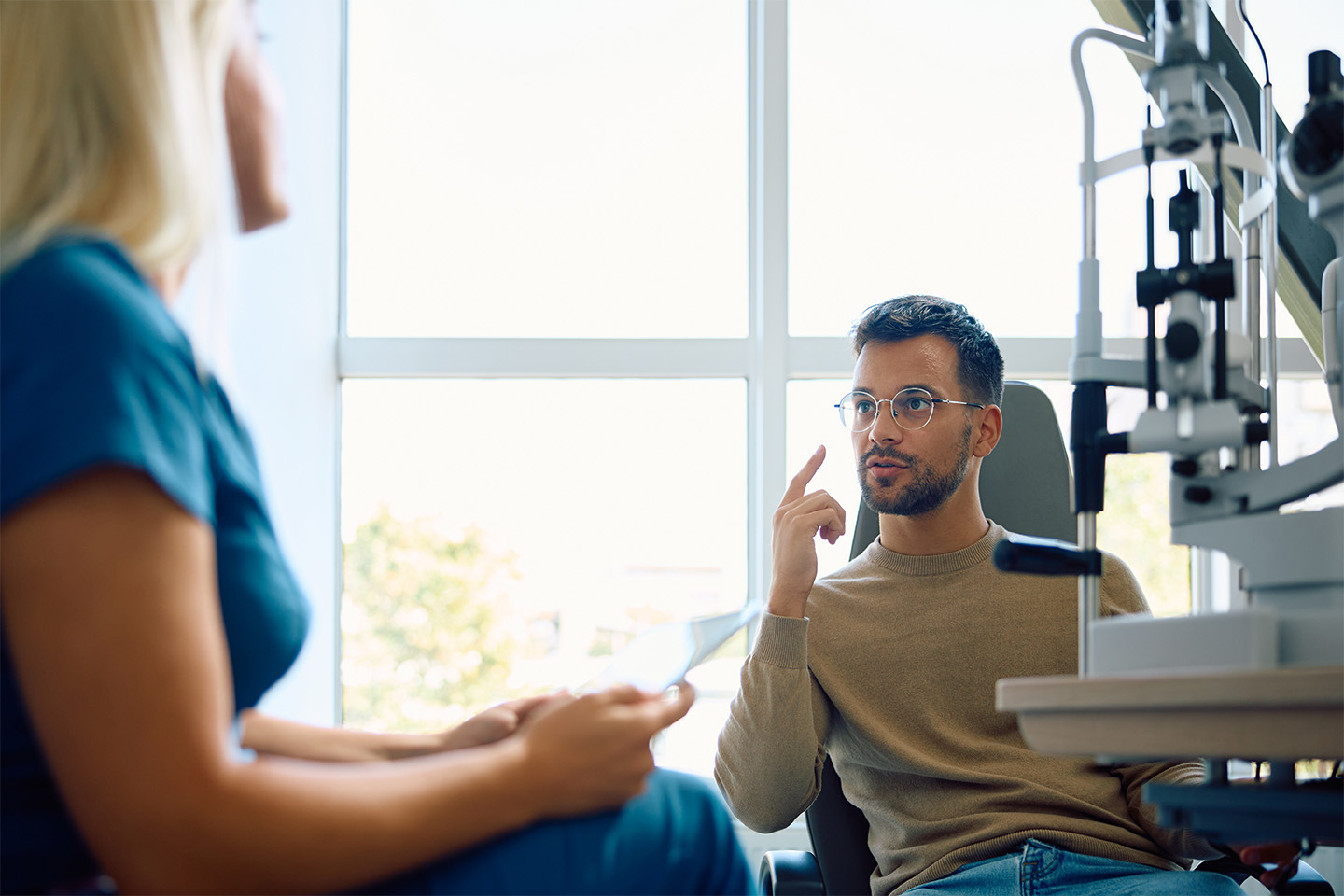
Getting effective treatment for glaucoma starts with getting the proper diagnosis. And at Kleiman Evangelista Eye Centers, the top eye doctors in greater Dallas employ several tests to determine the stage, form, and severity of your condition and work with you to make sure you understand your tests and what they mean. We’ll then work together to create a plan to keep your vision at its best.