Refer a Patient
 Pay Bill
 Patient Portal
Retinal Detachment Symptoms and Treatment
You never want to risk losing your eyesight. Unfortunately, certain conditions can quickly steal your vision and require immediate attention. Retinal detachment is one such condition that is considered an emergency. So, recognizing the early warning signs is vital to receiving swift and complete care and keeping your eyesight.
Continue reading to learn about the symptoms and treatment options for retinal detachment.
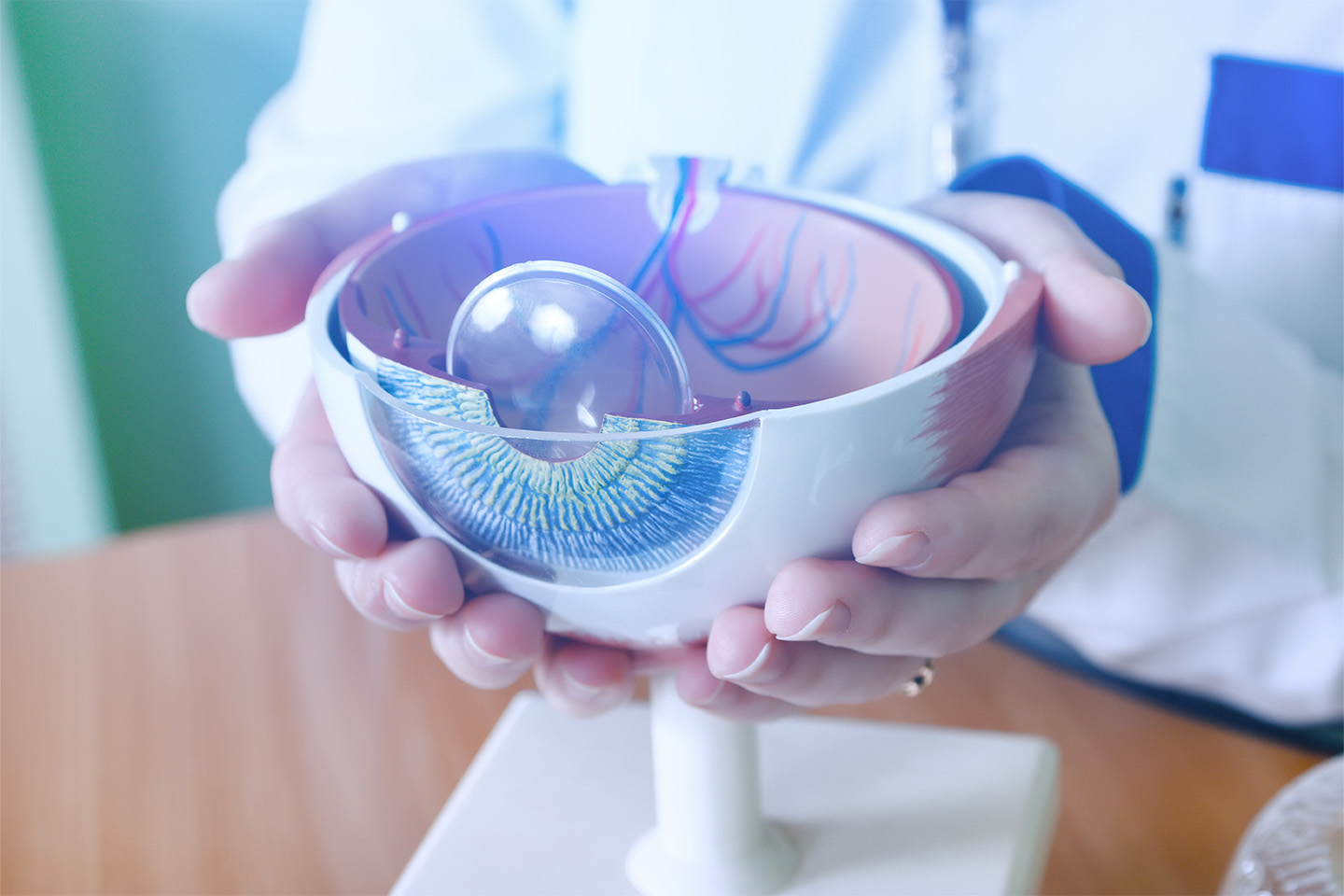
What is Retinal Detachment?
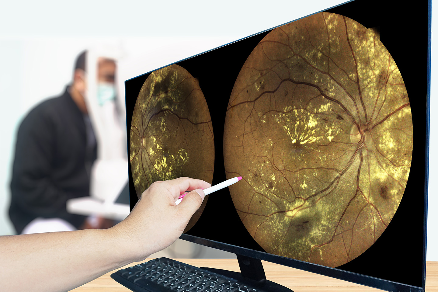
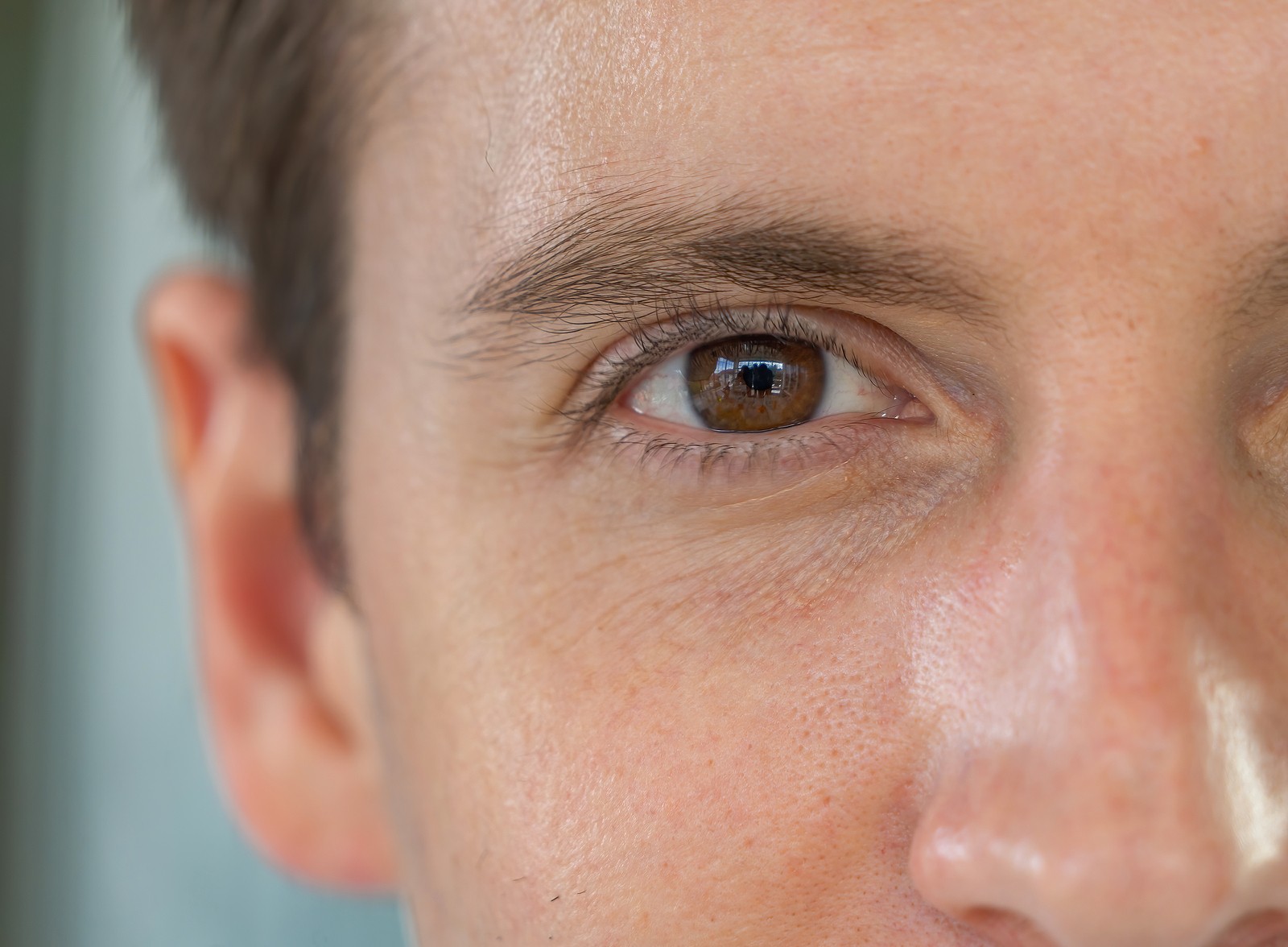
Retinal detachment occurs when the retina — a thin layer of tissue that senses light and sends signals to the brain — pulls away from the eye wall. A build-up of fluid between the wall and the retina can push it away. This condition separates photoreceptor cells from the back of the eye. If left untreated, it can cause permanent vision loss. The earlier it is detected, the better the chance of visual recovery.
Some characteristics can leave you more at risk for retinal detachment:
- Aging, especially those over 50
- Diabetes
- Age-related macular degeneration
- Eye injuries
- Inflammation disorders
- Extreme nearsightedness
- Surgery, including cataract removal
- Family history
Symptoms of Retinal Detachment
It is crucial to know the warning signs of retinal detachment and seek help as soon as you notice changes, including:
- Increased and sudden appearance of tiny moving specks (floaters) and flashes of light
- Decreased peripheral vision
- Blurry vision
- Reduced vision
- Poor vision in reduced light
- Curtain-like shadow over your visual field
Treatment for Retinal Detachment in Greater Dallas
Surgery is the most common solution for retinal detachment and should occur within days of your diagnosis to preserve your vision. Depending on whether you have a hole, tear, or total detachment, our ophthalmologists in the greater Dallas area will recommend the best treatment option.
1. Cryopexy
Cryopexy is a type of extreme cold therapy or freezing that creates scar tissue to seal tears and secure a small portion of the retina in place.
2. Laser Photocoagulation
Laser photocoagulation involves the creation of small burns on the retinal tissue to scar down holes or tears. This procedure is most commonly used for small tears or holes, but may be used to seal off a portion of the retina if the detachment is small.
3. Pneumatic Retinopexy
This treatment involves injecting an air or gas bubble into the center of the eye. The bubble pushes the retina against the wall of the eye to stop fluid from entering that space. Cryopexy is then used to secure the retina to the wall and allow for proper healing. Over time, fluid fills the eye and replaces the bubble. This is an in-office procedure and only possible if there is only one tear on the retina.
4. Scleral Buckling
Scleral buckling is when your surgeon sews a rubber, plastic, or silicone to the white of the eye to push it inward and let the retina heal against the eye wall. It typically remains in place permanently. This option is generally used on younger patients (under the age of 50) since it reduces the risk of developing an early cataract.
5. Vitrectomy
During this procedure, your surgeon removes the vitreous fluid and tissue that pulls on the retina. They will then replace it with an air or gas bubble to push the retina back into place. Additional laser barriers or cryopexy are typically used as well to seal any areas that are torn. This procedure often will lead to the development of a cataract if the patient has not already had cataract surgery.
What to Expect During Retinal Treatment Recovery
Recovery lasts for about 4 to 6 weeks, but it can take several months for your vision to improve. Some patients may never recover all lost vision. This is the case if the detachment goes through the very center of the retina, the macula. In some cases, a second surgery may be necessary for complete treatment. For the best results, you must follow specific aftercare instructions to ensure your eyes heal correctly.
- You may experience some discomfort during your recovery, but pain medication and eye drops can ease this.
- You’ll be expected to rest for weeks following your surgery, and your ophthalmologist will tell you when to resume driving, working out, etc.
- Keep your eye patch on for the recommended amount of time.
- You will be expected to keep your head in a specific position, so the bubble stays in place.
- If you receive an air or gas bubble, avoid flights, high altitude, or scuba diving, which can all impact the eye pressure. You must avoid these activities until the bubble dissolves completely.
As the experts in cataract and LASIK surgery in greater Dallas, we aim to help you maintain the best vision possible with a range of tailored surgical solutions. If you suspect that you are experiencing retinal detachment, schedule an appointment as soon as possible to preserve your vision.
[DISPLAY_ULTIMATE_SOCIAL_ICONS]